
Norovirus conformational changes may provide insights into novel therapies
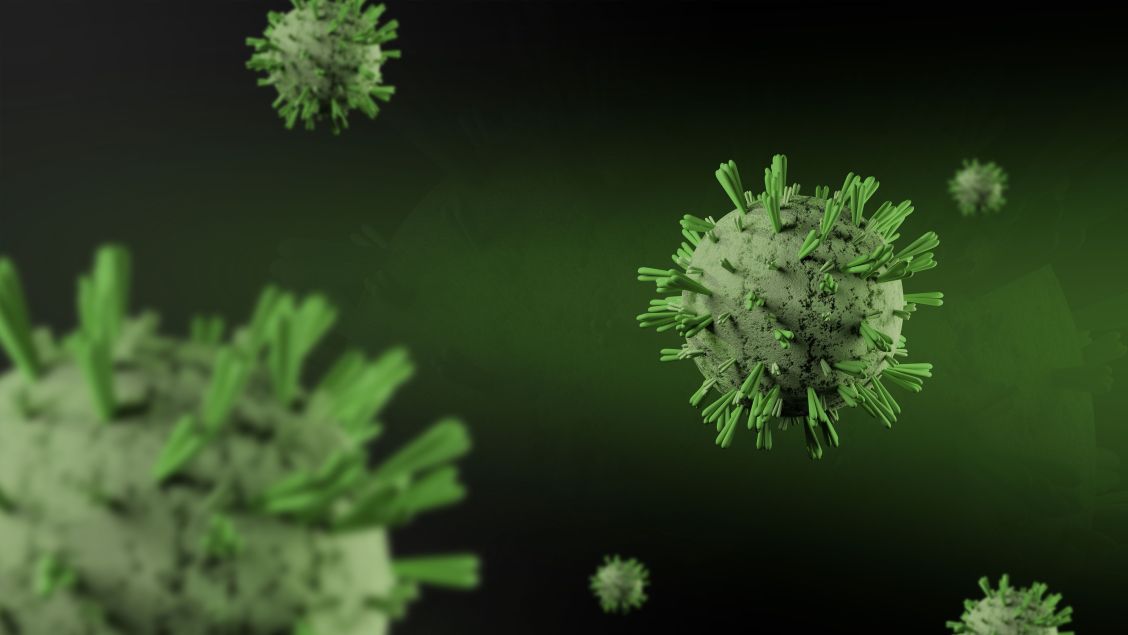
Human norovirus (HuNoV) is a leading cause of acute gastroenteritis worldwide, causing an estimated 600-700 million infections and almost 200,000 deaths each year.
Yet, despite its significant impact, there are currently no effective antiviral treatments for HuNoV. Understanding how the virus interacts with host cells and the immune system is therefore important for developing new ways to prevent and treat infection.
One challenge for researchers is that HuNoV is difficult to grow using conventional cell culture systems. This has made virus-like particles (VLPs) an important tool for studying the virus. VLPs closely resemble the outer structure of norovirus but are much safer as they lack infectivity and cannot replicate.
The outer shell of the virus, called the capsid, plays an important role in norovirus infection and immune recognition, with regions being involved in interactions with host receptors and antibodies. Understanding how this structure changes could highlight how the virus infects cells and how new vaccines or antiviral treatments could be developed.
Previous studies on murine norovirus, a variant which infects mice, demonstrated that the virus has a dynamic structure capable of undergoing conformational changes from a resting state to a rising state.
A new study, published in the Open Access journal International Journal of Molecular Sciences (IJMS), presents the first direct structural evidence for conformational changes in human norovirus. Using cryo-electron microscopy (cryo-EM), the structure of the HuNoV VLPs were characterised and two distinct conformations were identified corresponding to resting and rising states. The findings provide new insight into the structural flexibility of HuNoV and could help to inform future vaccine design and antiviral development.
A closer look at the norovirus capsid
HuNoV is composed of two separate proteins:
- VP1: The major capsid protein which makes up most of the virus shell and is responsible for receptor binding and is the region which the immune system recognises.
- VP2: The minor structural protein which is located inside the capsid where it helps to stabilise the structure.
The norovirus capsid is made up of 180 copies of VP1 arranged into an icosahedral structure. Each VP1 subunit contains a shell domain (S), which forms the main capsid structure, and a protruding (P) domain that extends from its surface.
The P domain contains two subdomains, P1 and P2. The P2 subdomain is particularly important because it contains regions involved in interactions with host receptors and is also a major target for antibodies. Unlike the relatively conserved S domain, the exposed P2 region varies between norovirus strains and is associated with immune escape mechanisms. Changes in the shape of the P domain could provide another way for the virus to alter how these regions are recognised by the immune system, by changing which regions are exposed to antibodies.
How the virus regulates receptor engagement and immune recognition is not fully understood. However, previous studies of murine norovirus, which infects mice, have shown that the P domain can move in response to environmental conditions such as pH and metal ions. In these viruses, the domain can adopt a resting position close to the capsid shell or a raised position further away from it. These movements change the position of the P2 subdomain on the capsid surface, potentially altering how accessible its receptor and antibody binding regions are. However, direct structural evidence for similar conformational changes in human norovirus has been limited.
Discovering HuNoV capsid exists in two forms
To investigate whether HuNoV could undergo similar changes, the researchers produced VLPs using a baculovirus expression system, which allows the production of large amounts of proteins inside cultured insect cells. They then used cryo-EM (Figures 1A, 1B) to image the particles and analyse their structures.
The researchers found a group of particles, approximately 40 nanometres in diameter, which existed in two distinct structural populations:
- Resting state: The P domains remained close to the S shell (Figure 1C).
- Rising state: The P domains were elevated away from the S shell (Figure 1D).
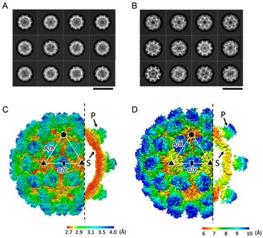
Figure 1: A) Cryo-EM structure of HuNoV capsids in the resting state. B) Cryo-EM structure of the HuNoV capsids in the rising state. C) Map of the protein structure in the resting state with the P and S domains annotated. D) Map of the protein structure in the rising state with the P and S domains annotated.
The rest of the capsid structures appear identical, with the main differences appearing from a 55° rotation of the P domain and it moving between 1.0 and 1.3 nanometres away from the shell.
The rising state also displayed higher flexibility. Analysis showed that the position of the P domain in this state could vary by around 11 Å between different particles, which was compared to smaller movements in the resting state. This greater flexibility made the domain more difficult to resolve using cryo-EM, giving a final resolution of 4.7 Å for the P domain. While resolutions below 2 Å are generally considered near atomic, resolution can be affected by factors such as the size and complexity of a protein and the flexibility of individual regions.
Importantly, both conformations were discovered in the same purified VLP sample, produced under identical conditions. This suggests that in reality, both states exist in an equilibrium and that differences in conformation are not the result of sample differences but do represent examples of structural variation within the HuNoV capsid itself.
Changing the connections between VP1 proteins
The movement of the P domain also changed how neighbouring VP1 proteins interacted.
In the resting state, neighbouring P proteins formed contacts primarily through the P2 domain. The interactions helped to keep P domains close together and maintain the compact arrangement of the capsid surface.
When the P domains moved into the rising state, these P2-mediated interactions were disrupted. Instead, interactions shifted towards the P1 subdomain.
The researchers found that some of the amino-acid residues involved in these interactions vary between different strains of human norovirus. Variation in these regions could alter the interactions that stabilise the rising and resting states, potentially shifting the equilibrium between the two conformations. This could affect the structure and accessibility of the P domain, helping to explain why different norovirus variants can have different properties, including differences in receptor interactions, infectivity, and immune recognition.
Could changing shape change infection?
The structural changes observed in this study, notably the GII.3 strain of the virus, could have consequences for how norovirus interacts with host cells and the immune system.
The structural changes observed in GII.3 could affect how the virus interacts with host cells and the immune system. The P2 subdomain contains regions involved in binding host histo-blood group antigens (HBGAs). Raising the P domain changes the position and orientation of the P2 surface, which could alter how these regions interact with receptors and antibodies.
Previous research shows that an antibody can bind to the norovirus without necessarily stopping infection, suggesting that where an antibody binds is not the only factor that determines whether it can stop infection. The shape and stability of the capsid may also affect how well an antibody can neutralise the virus.
The new structures provide a possible explanation. The amino-acid differences found between norovirus strains could affect the interactions that hold the capsid in its resting or rising state. This could change how often the virus adopts each shape, and therefore how accessible parts of the P domain are to antibodies. Differences in this balance could help explain why an antibody can neutralise one norovirus variant but not another.
Could these results aid the design of a vaccine for human norovirus?
The discovery of two GII.3 capsid conformations adds to evidence that norovirus capsids are dynamic rather than fixed structures.
However, several questions remain. The researchers identified both states under the conditions used for their experiments, but they did not provide an insight into what causes the virus to adopt one state over the other. External factors that change throughout the body, such as pH, temperature, and oxygen and salt levels could affect the balance between these two conformations, however, these were not tested in this study.
Understanding what controls this structural switch could help researchers determine whether the two states have different roles during infection. Previous work in murine norovirus has linked capsid conformational changes to receptor binding, infectivity, and immune evasion, but the biological consequences of the rising state in human norovirus (HuNoV) remain uncertain.
The findings could have a future impact on vaccine and antiviral development. Because the P2 surface contains important receptor-binding and antigenic regions, changes in its position and orientation could affect which parts of the capsid are exposed to the immune system. Targeting this conformational behaviour of the capsid, rather than a fixed structure, could therefore provide another approach for developing treatments or vaccines.
Ultimately, this study provides a new structural picture of the GII.3 HuNoV capsid, where the protruding P domains can move and the surface of the virus can take more than one form. Further work will be needed to establish what drives these changes and what they mean for infection, but the discovery provides a useful starting point for studying how the structure of HuNoV changes during its lifecycle.
More research on virology, structural biology, and infectious disease can be found across MDPI’s Open Access journals, Biology, Antibiotics, and International Journal of Molecular Sciences.
Alternatively, you can access the full MDPI journal list here.