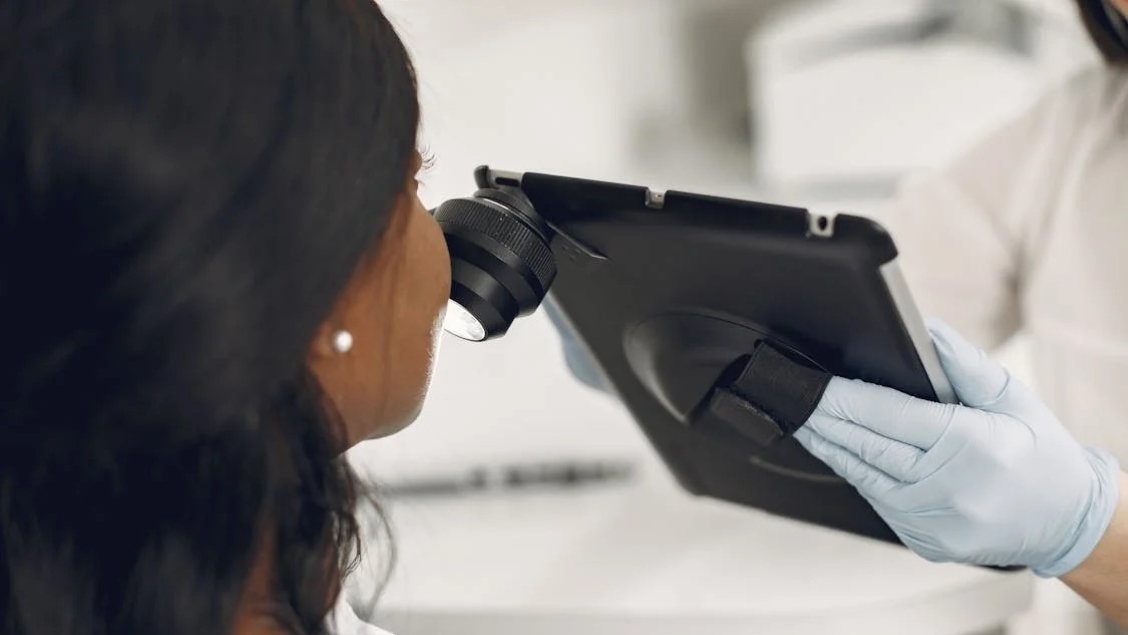
Exploring AI for Skin Diagnostics
The use of artificial intelligence (AI) in the healthcare industry has accelerated in recent years as the technology has become increasingly sophisticated. It is used in various settings such as screening, diagnostics, data handling, and for predictors of prognoses. A review published in the Open Access journal Life explores the use of AI in fluorescence photography (FP) in the context of skin assessments.
Fluorescent photography is a brilliant high-contrast imaging tool that can be used as a non-invasive technique for assessing human skin pathologies. It is also useful for cosmetics and other types of dermatological research. Skin analysis specialist researchers from Haut.AI explore AI, and how this powerful technology can be utilised with FP to generate highly detailed skin analyses. This review also explores the intricacies of the FP method and its applications in dermatology.
How does fluorescent photography work?
Fluorescent photography works by highlighting fluorophores present in skin cells which have different levels of light emission. Therefore, upon exposure to ultraviolet light, the skin fluorophores exhibit various intensities which are then captured in a photograph. These images are analysed, and a skin analysis can be carried out to see if there are any pathologies such as acne, aging or hyperpigmentation.
The comprehensive review discusses the main uses of FP, which include the assessment of photoaging through the detection of dark spots and pigmentation. Furthermore, FP remains an important part of diagnosing skin malignancies and is useful for the early detection of skin cell irregularities.
For this type of tool integrating AI for the analysis could be highly beneficial for skin diagnostics, and determining pathologies as well as cosmetology.
Fluorescence photography allows us to see what the human eye often cannot, and when combined with AI, we’re unlocking entirely new levels of skin diagnostics. – Anastasia Georgievskaya, CEO and co-founder of Haut. AI.
Artificial Intelligence (AI) for skin diagnostics
Detecting the early onset of photoaging and acne could be highly useful in determining efficient treatment plans to reduce the progression of skin abnormalities. The review describes previous studies showing how AI algorithms and deep neural networks can classify areas of skin with acne, inflammatory lesions and other skin abnormalities from the analysis of bright field images.
These AI-powered algorithms can be used in combination with other technologies. These technologies have been incorporated in wearable devices and mobile app tracking has been used for the early detection of abnormalities and continuous monitoring. Furthermore, the use of this innovative technology can be beneficial in reducing in-person visits to doctors for skin assessments.
Combining AI with fluorescent photography
Unlike FP, bight-field imaging is a technique that is routinely used for diagnostic purposes in skincare and dermatology. The process involves taking a skin sample and analysing the cellular structures, components and lesions under a standard light microscope.
With FP providing much more specific and detailed imaging compared to bright-field imaging, it could be extremely useful to integrate AI with FP for analysis, diagnostics, and potential development of treatment plans. The authors discuss how this could generate a highly refined analysis of the skin.
The ethical implications of using the tool are also discussed in the review. Using AI systems poses potential ethical issues regarding privacy and access to personal biodata. There are currently no global policies in place to regulate the use of AI and its access to patient data in the fields of dermatology and cosmetology. The authors state that safe storage of biodata and adherence to legal policy are crucial to ensure the ethical incorporation of AI-powered analysis of patient data in all aspects of
Further research
FP has proven to be a useful tool in detecting skin abnormalities through contrasting levels of light emitted by skin fluorophores. This includes the detection of issues such as acne, hyperpigmentation and skin malignancies. Bright-field imaging techniques have previously been used in tandem with AI algorithms, but not with FP, which offers more specific and detailed images of cellular structures in the skin.
We’re looking at a future where skin analysis is multimodal and utilizes different ageing models and biomarkers, such as using fluorescence spectroscopy, to make skin analysis more precise and predictive. – Anastasia Georgievskaya
There are currently no datasets for the use of FP in detecting skin malignancies, which would be vital to integrating AI algorithms with the imaging tool. This review provides information on the mechanics of FP and the applications of AI in skin diagnostics. Importantly it provides a discussion on how it can be integrated with FP to potentially revolutionise the current landscape of skin diagnostics.
Read more about this research by accessing articles published in the Open Access journal Life or access the full list of MDPI journals.


