Exciting New Tools in Breast Cancer Diagnostics
To wrap up October and Breast Cancer Awareness Month, we’re exploring some of the exciting advancements being made in breast cancer research. This is made possible by the dedicated minds and progress made by researchers and healthcare workers in oncology.
There are a staggering 55,920 breast cancer each year in the UK alone. Breast cancer is the most common form of cancer that women are diagnosed with, but both men and women can get breast cancer. Seventy-six percent of people diagnosed survive for 10 years or more, with survival based on a variety of factors (including age, sex, and type of cancer).
Oncologists, assess, diagnose and treat people with cancer. People with breast cancer that is detected early and has not spread, or metastasized, have much higher chances of survival. This highlights the importance and need for accurate and efficient diagnostics. Here we introduce the use of imaging in breast cancer. Furthermore, we explore the recent advancements, including ways in which artificial intelligence and liquid biopsies can be used in the clinic.
Mammograms
Multiple diagnostic tools have been developed and are now clinically used to determine the presence of tumours and tumour types. Mammograms are the most common imaging tool currently used in the clinic.
Mammograms are essential for the early detection of breast cancer. In the UK, females from the age of 53 (higher-risk individuals), are prompted to take a mammogram. This national screening program has been instrumental in catching the disease early, and therefore, in saving lives.
2D mammograms were introduced for breast cancer in the 1960s. They work by taking X-ray images of the front and side of the breast, and then putting them together into one image.
Then in 2008, 3D mammograms were introduced, with this advanced technology differing slightly by taking multiple X-ray images from different angles and then combining those images together. As the name suggests, it formulates a 3D image of the breast and yields much higher clarity and a more efficient way of locating tumours. Studies have shown that women undertaking 3D mammograms had better outcomes, and may also yield fewer false positives compared to 2D mammograms.
False positives and negatives
But what are false positives? False positives occur when the presence of a tumour is suspected by radiologists in a mammography image, but then follow-up tests reveal normal breast tissue instead. These false positives are very common when taking a mammogram, exhausting time and resources, not to mention increase the anxiety of patients waiting for test results. Similarly, but much more detrimental, false negatives can also occur, indicating no presence of a tumour when there is in fact cancer present. However, this is much more rare. Scientists continue to look for more efficient ways to reduce the issue of human error when reading scans. This is where artificial intelligence (AI) comes in.
Using artificial intelligence for diagnosis
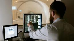
With the use of AI in medical research catapulting in the last decade, it’s exciting to see its incorporation into cancer research. One of the ways scientists are trying to use these complex learning algorithms is to accurately detect tumours from mammogram images. AI can detect 20% more cancers from mammograms than without AI. The use of AI in reading mammograms could drastically speed up and improve breast cancer diagnosis, improve clinical resources, ease patient waiting time and in turn decrease patient anxieties.
Another way scientists are using AI in breast cancer diagnostics is explored in a study published in Cancers. The clinical study aimed to classify lymph nodes of early-stage breast cancer patients using AI in PET scanning. They created a deep-learning AI model which analyses the lymph nodes in PET images. This analysis would then assist oncologists in determining the type of breast cancer and treatment plan of the patients.
Deep learning in breast cancer continues to remain a hot topic in the field of breast cancer research. We hope to see the future application of this exceptional technology in the clinic to further assist oncologists oncologists to make the right calls in breast cancer diagnostics and treatment.
Liquid Biopsies
Moving away from imaging, we turn to liquid biopsies, a novel way to detect the presence of cancer, and an exciting area of cancer research.
Liquid biopsies are a non-invasive technique usually used alongside initial scans to confirm the presence of a tumour. They detect the presence of cancer-specific components, known as ‘markers’, in the blood or saliva, including circulating tumour cells (CTC), and the DNA released from these cells (ctDNA). The biopsy is therefore able to give the doctor more information on the specific type and stage of breast cancer. Hence, this method is potentially an excellent way to determine the type of treatment the patient will receive.
Liquid biopsies are not only used during the diagnosis of breast cancer but are also increasingly used to monitor patients during treatment and after treatment. The tool can be used to measure how the body reacts to the drugs and to test for recurrence of cancer after treatments have finished.
Liquid biopsies and breast cancer
A Diagnostics review discusses the current progressions of liquid biopsies and its use in the clinic. As mentioned, liquid biopsies are commonly used to assess the response to treatment. However, the review also addresses current limitations including its difficulty in detecting breast cancer in its earlier stages. Hence, clear markers of early-stage breast cancer are needed to maximise the efficiency of using liquid biopsies.
Researchers are still finding different ways to utilise this powerful technique. Specific components (such as ctDNA and CTC) can be harnessed through liquid biopsies. Therefore, analysing the features of these components could be used as potential biomarkers. This idea is explored in a study published in Cancers, which analyses two different cell types obtained from liquid biopsies. They found that the presence of these different cell types in the blood may be associated with cancer metastasis and progression.
Detecting markers for breast cancer prognosis is one of the many potential applications of liquid biopsies that we hope to see make its way into future clinical practice.
Future research in breast cancer
These new tools propose an exciting new era for breast cancer diagnostics, with scientists aiming to refine the current methods in the clinic.
You can read more about this exciting area of research in Cancers, Current Oncology and Diagnostics. These MDPI Journals explore some of the latest ideas and topics in the field of oncology and diagnostics. If you would like to submit your own research, please see the full list of MDPI’s Open Access journals for more information.


