Circulating Tumor Cells: An Interview
Circulating tumor cells intravasate and extravasate (enter and exit the blood vessels) allowing migration to other parts of the body. Tumor metastases cause ninety percent of cancer deaths. Metastases occurswhen cells detach from a primary tumor and form new malignancies elsewhere.
Recent technological developments enable detection of minute amounts of circulating tumor cells in patient blood samples. These non-invasive “liquid biopsy” techniques allow patient monitoring and early detection of metastasis, and provide the possibility for cell-specific information to direct personalized, targeted therapy at an early stage of the disease.
In Towards the Biological Understanding of CTC: Capture Technologies, Definitions and Potential to Create Metastasis, Barradas and Terstappen review the current state of CTC research and technology in a highly informative work recently published in the journal Cancers Special Issue Circulating Tumor Cells in Cancer. Below, Leon Terstappen, discusses challenges and directions in CTC research and technology.
An interview with Leon Terstappen about circulating tumor cell technologies
We were privileged to have a chance to sit down and talk with Leon Terstappen. We learned a great deal about CTC capture technologies.
On the definition of circulating tumor cells for diagnostic and therapeutic values for CTC capture technologies
LT: Each technology is using a different definition. The most common technologies are chasing the EpCAM (Epithelial Cell Adhesion Molecule) antigen, at least for carcinomas, so that represents a large camp of technologies, but even within that camp there is enormous discrepancy because how do they then identify the cells? Many are doing it with microscopy technologies but even there, what is the numerical aperture of the objective, which reagents and colors do you use to identify the cells?
The standards are basically from the CellSearch system using a nucleic acid dye (DAPI), using antibodies against some of the cytokeratins, labeled with PE, and CD45, a molecule expressed on leukocytes labeled with APC. So then the original definition is that it has to express cytokeratins, lack CD45, be at least 4 microns and look like a cell, which is of course, subjective .
When you change any of the dyes or the reagents, the cells which you will be counting will not be the same cells anymore, but how do you know what is the correct definition? There are reports of people detecting a thousand times more circulating tumor cells, but then you are in the range of the white blood cells, so if that were so, everyone should have seen them. Sometimes detection might include tumor microparticles, not really cells, since a lot of technologies do not allow you to actually see that. As the technologies develop, the number of detected cells goes down because you add criteria, which makes it more strict.
The challenge for investigators
LT: So the real challenge here is to have the various investigators use a common platform at least to share definitions or let other people look at cells and say this is one, that’s not one, otherwise you never get consensus, so in the end, you will have to do a clinical study to demonstrate that the presence of these cells indeed strongly correlates with the clinical outcome. The CTCs detected by the CellSearch system have been proven in clinical trials in that the more CTC you have the quicker you die. When you suddenly find ten times more, also in patients where you didn’t see them with the CellSearch system, that is confusing because these patients live much longer. So, perhaps then the cells, if they are in fact cells, which you are detecting, are not really relevant.
Explaining current circulating tumor cell research as well as CTCtrap’s role in advancements
LT: The technologies to reliably capture CTC and investigate them are fairly recent and allow us to gather information on what you see in these cells at the protein and gene level. But the ability to investigate heterogeneity means also you have to do so at the single cell level, so we are currently developing novel technologies to actually enable that because the most critical aspect in cancers is the heterogeneity and the constant changing. You are diagnosed today with metastatic disease and you get treated. But the phenotype and genotype of the tumor cells change and they will become resistant to the therapy. These cell specific investigations are proceeding currently, but will progress faster if most people would at least have similar definitions about what they call a CTC, so that then in reports about gene expression, protein expression, you are talking about the same thing.
An exciting future for the technology
LT: The real promise of this technology is to personalize therapy, and the only way to do that is to have tumor cells available at all times during the disease process, so that you can actually change therapy when needed according to the new information. The information to direct therapy options is contained within these cells, so we hope to get as much information out as possible and hopefully in the future we can treat the patients based on the phenotype and genotype of the tumor cell that is actually circulating in the patient’s blood.
About Professor Terstappen
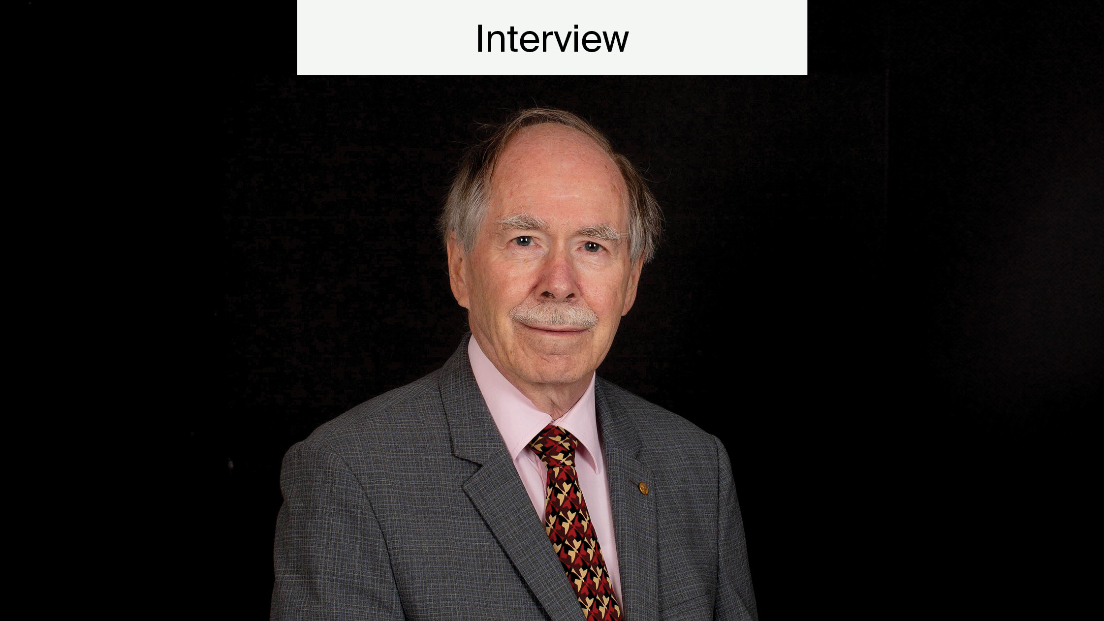
Professor Leon Terstappen of the University Twente has been particularly successful at developing innovative technology into practical clinical application. He is responsible for the development of CellSearch, the only FDA-approved CTC capture technology. In 2009 he was awarded the highly prestigious Prix Galien for this endeavour. He also initiated the development of the CTCtrap (Circulating Tumour Cells TheRapeutic Apheresis) project. This was a six million euro, EU-funded international effort, to filter entire patient blood volumes for CTC detection and analysis.
Leon Terstappen received his Medical Degree from the University of Groningen in 1983. He received his PhD from the Department of Applied Physics of Twente University in 1987. In addition, he worked on characterization of hematopoiesis as a post-doctoral fellow and in various research positions at the research institute of Becton Dickinson, San Jose, CA, USA. Professor Terstappen joined Immunicon Corporation in 1994 to pioneer the detection of tumor cells in blood of cancer patients, eventually becoming Chief Scientific Officer and Senior Vice President of Research and Development. He was appointed Professor of Medical Cell Biophysics of the University of Twente in 2007. Professor Terstapen is a consultant of Veridex LLC and receives research funding from Veridex LLC. More information can be found on his webpage.
References
Uhr, J.W. The Clinical Potential of Circulating Tumor Cells; The Need to Incorporate a Modern “Immunological Cocktail” in the Assay. Cancers 2013, 5, 1739-1747.
Galanzha, E.I.; Zharov, V.P. Circulating Tumor Cell Detection and Capture by Photoacoustic Flow Cytometry in Vivo and ex Vivo. Cancers 2013, 5, 1691-1738.
Hu, B.; Rochefort, H.; Goldkorn, A. Circulating Tumor Cells in Prostate Cancer. Cancers 2013, 5, 1676-1690.
Charpentier, M.; Martin, S. Interplay of Stem Cell Characteristics, EMT, and Microtentacles in Circulating Breast Tumor Cells. Cancers 2013, 5, 1545-1565.
Markiewicz, A.; Książkiewicz, M.; Seroczyńska, B.; Skokowski, J.; Szade, J.; Wełnicka-Jaśkiewicz, M.; Zaczek, A.J. Heterogeneity of Mesenchymal Markers Expression—Molecular Profiles of Cancer Cells Disseminated by Lymphatic and Hematogenous Routes in Breast Cancer. Cancers 2013, 5, 1485-1503.
Andergassen, U.; Kölbl, A.C.; Hutter, S.; Friese, K.; Jeschke, U. Detection of Circulating Tumour Cells from Blood of Breast Cancer Patients via RT-qPCR. Cancers 2013, 5, 1212-1220.


